

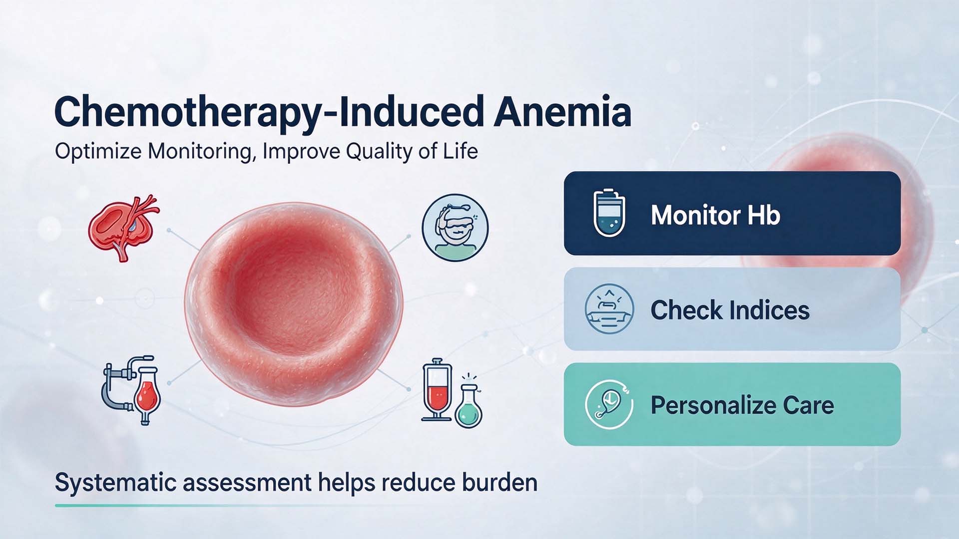
Chemotherapy-induced anemia (CIA) is a common, debilitating complication affecting up to 70% of cancer patients undergoing myelosuppressive treatment. Characterized by reduced red blood cell production secondary to chemotherapeutic agents’ toxic effects on bone marrow erythroid progenitors, CIA is associated with fatigue, impaired quality of life, reduced treatment tolerance, and even poorer oncological outcomes. A systematic approach to interpreting red blood cell parameters, combined with standardized monitoring protocols, is critical to minimizing the burden of this condition.
The core diagnostic features of CIA align with well-established hematological principles: red blood cell (RBC) count, hemoglobin (Hb), and hematocrit (Hct) typically decrease in parallel, as chemotherapy suppresses overall erythropoiesis. These three parameters are intrinsically linked by hematological formulas: Hct approximates the product of RBC count and mean corpuscular volume (MCV), while Hb equals RBC count multiplied by mean corpuscular hemoglobin (MCH). In many cases, CIA presents as normocytic normochromic anemia, with normal MCV and MCH values, though patients with concurrent nutritional deficiencies may show microcytic or macrocytic features. It is important to distinguish CIA from other causes of anemia in cancer patients, such as chronic bleeding, iron deficiency, or bone marrow infiltration by tumor cells: isolated Hb reduction with normal RBC count often indicates occult gastrointestinal bleeding, while elevated Hct with normal Hb may stem from dehydration caused by chemotherapy-induced nausea and vomiting.
Accurate interpretation of these parameters relies on three core validation rules. First, numerical consistency must be verified: the calculated Hct (RBC × MCV) should align with the measured Hct, with discrepancies exceeding 3% indicating potential pre-analytical errors such as specimen hemolysis or clotting, a common issue in oncology patient samples. Second, unit conversion pitfalls should be avoided: while Hb is measured in g/L in most domestic systems, international laboratories may report values in g/dL, requiring careful conversion to avoid misclassification of anemia severity. Third, parameters should always be interpreted alongside MCV, MCH, and mean corpuscular hemoglobin concentration (MCHC) to identify concurrent nutritional deficiencies that may exacerbate CIA.
Hemoglobin testing forms the cornerstone of CIA monitoring, with clinical guidelines recommending testing before each chemotherapy cycle to assess anemia severity and guide intervention. For patients receiving erythropoiesis-stimulating agents (ESAs), the mainstay of CIA treatment, regular hemoglobin testing every 2–4 weeks is mandatory to adjust dosage, ensuring Hb levels remain within the target range of 100–120 g/L; levels exceeding 120 g/L are associated with a 30% increased risk of thromboembolic events, a particularly dangerous complication in cancer patients. For patients with severe CIA requiring transfusion support, hemoglobin testing prior to each transfusion helps avoid unnecessary blood product use and reduces the risk of transfusion-related adverse events.
Hemoglobin testing machine forms the cornerstone of CIA monitoring, with clinical guidelines recommending testing before each chemotherapy cycle to assess anemia severity and guide intervention. For patients receiving erythropoiesis-stimulating agents (ESAs), the mainstay of CIA treatment, regular hemoglobin test system every 2–4 weeks is mandatory to adjust dosage, ensuring Hb levels remain within the target range of 100–120 g/L; levels exceeding 120 g/L are associated with a 30% increased risk of thromboembolic events, a particularly dangerous complication in cancer patients. For patients with severe CIA requiring transfusion support, hemoglobin testing prior to each transfusion helps avoid unnecessary blood product use and reduces the risk of transfusion-related adverse events.
When abnormal red cell indices are identified, a standardized management workflow should be followed. First, verify the logical consistency of parameters to rule out laboratory errors. Second, correlate results with clinical context: recent chemotherapy cycles confirm myelosuppression as the likely cause, while symptoms such as melena require investigation for gastrointestinal bleeding. Third, order supplementary tests as needed, including iron studies, vitamin B12 levels, and reticulocyte count to assess bone marrow erythropoietic activity. Finally, implement personalized intervention: ESAs are indicated for moderate anemia, while transfusion is recommended for Hb <70 g/L or symptomatic severe anemia. With systematic monitoring, including regular hemoglobin testing, clinicians can effectively manage CIA, improve patient quality of life, and ensure uninterrupted delivery of life-saving cancer treatment.
Post time: Nov-22-2026