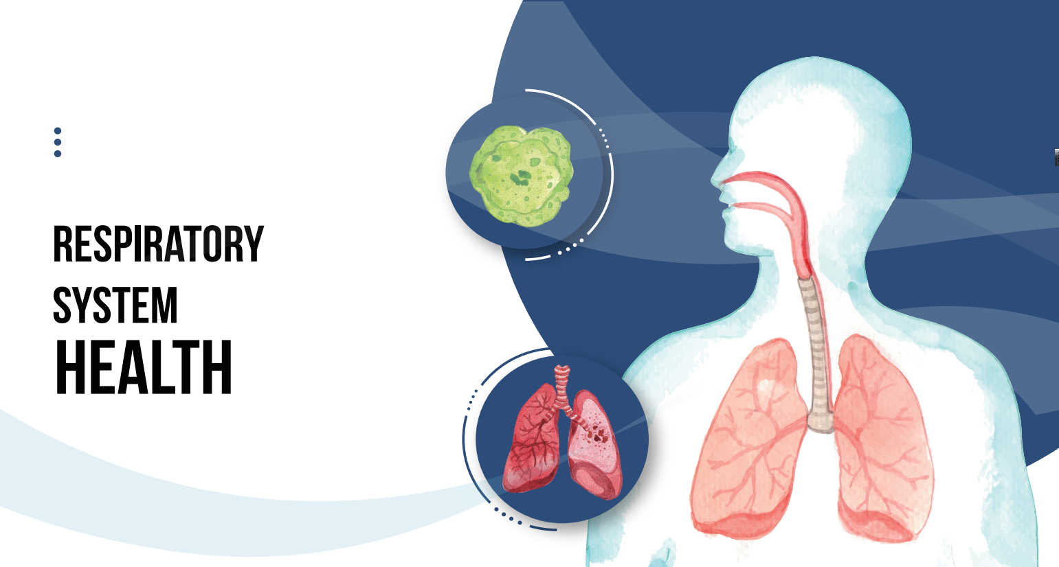
The respiratory tract of the human body is connected to the outside world, and the types of infectious pathogens are very complex, covering bacterial, fungal, viral, and even parasitic infections. Lower respiratory tract infection (LRTI) is one of the most common infectious diseases in the world. Pneumonia caused by respiratory tract infections is a significant cause of morbidity and mortality in the population, and typically has a significant impact on the elderly and immunocompromised population. Although there are multiple laboratory diagnostic methods available, about 40% of community-acquired pneumonia (CAP) patients who require hospitalization still have difficulty diagnosing the pathogenic pathogen. Therefore, improving the accuracy and effectiveness of laboratory diagnosis is of great significance. Traditional detection methods, including cultivation, are still considered the “gold standard” for pathogen detection while being improved. The development of molecular biology diagnostic techniques has been widely used in microbiological diagnosis of respiratory pathogens in recent years.
Development and clinical application status of pathogen detection technology for respiratory tract infections
1.1 Traditional respiratory pathogen detection methods
1.1.1 Smear staining microscopy (1) There are many staining methods, commonly used are Gram staining and acid fast staining. Gram staining can be used to distinguish between Gram positive cocci and Gram negative bacilli, making it easy to operate. There are reports that the sensitivity of the Gram staining method in detecting cases of coccal pneumonia is only about 80%, especially for infections of Streptococcus pneumoniae and Staphylococcus aureus in the CAP population. Acid fast staining can highlight slender rod-shaped bacteria stained red, with high diagnostic specificity (90% to 100%) for mycobacteria, but limited sensitivity. Other staining methods, such as ink staining, have high diagnostic specificity for Cryptococcus and are particularly suitable for staining Cryptococcus in cerebrospinal fluid samples. However, their sensitivity is poor and should be combined with the Cryptococcus capsule polysaccharide antigen test. Hexamine silver staining is suitable for staining classical fungi and Yersinia pneumoniae. The most commonly used diagnostic method for fungal infections in sputum and bronchoalveolar lavage fluid samples is fluorescence staining with fluorescein coupled monoclonal antibodies. (2) Microscopic observation of cellular pathological effects is an important means of virus identification through culture. If stained alkaline inclusion bodies are found in respiratory sample cells, it can be diagnosed as adenovirus infection. The pathological effects of infected cells include giant cells, formation of syncytial bodies, and intracellular inclusion bodies. Microscopic examination has high specificity for specific detection items, but it is time-consuming and labor-intensive. In recent years, as a clinical diagnostic experiment, it has gradually been replaced by molecular biology.
1.1.2 Antigen detection method
Refers to the method of using known pathogen antibodies to detect the presence of corresponding pathogen antigens in a patient’s body. Common operating methods include latex agglutination test, colloidal gold immunochromatography, direct fluorescence antibody test (DFA), etc. As the preferred method for rapid detection of influenza, it can detect influenza A virus, influenza B virus, respiratory syncytial virus, etc. It is convenient to use and has high specificity (90% to 95%), but low sensitivity (40% to 70%), which means that negative results cannot exclude influenza virus infection. DFA can detect adenoviruses, respiratory syncytial viruses, etc. with high specificity, but the interpretation of results may be subjective and require high professional requirements. The preferred diagnostic method for cryptococcal infection is to use serum and cerebrospinal fluid samples for latex agglutination test or enzyme-linked immunosorbent assay (ELISA) to detect cryptococcal capsule polysaccharide antigen. The detection of Legionella pneumonia can be diagnosed by combining urine Legionella antigen type I detection with sputum agar culture. A novel immunochromatographic method for the detection of pneumococcal antigen in urine is used for the diagnosis of pneumococcal pneumonia. It is worth noting that many patients have already started antibiotic treatment during sample testing, and it is likely that bacteria cannot be cultured in the sample, but antigens still exist in the body. Therefore, antigen detection methods make up for the shortcomings of staining microscopy and traditional culture methods in this regard.
1.1.3 Cultivation method
Phlegm samples and bronchoalveolar lavage fluid samples are generally inoculated and cultured on blood plates, chocolate plates, MacConkey plates, etc. If there is suspicion of bloodstream infection, the pathogen is cultured in a blood culture bottle. Special culture media can also be selected for different pathogenic microorganisms in clinical practice. Using BCYE selective culture media to cultivate respiratory secretions is the gold standard for detecting Legionella infection, with high specificity but low sensitivity (50% to 80%). In addition, BCYE can also be used for the cultivation of Nocardia. The most definitive method for diagnosing invasive aspergillosis is to obtain positive culture results from normal sterile sites, usually using Sabouraud medium, but with lower sensitivity.
1.1.4 Serological identification
It refers to the use of immunological testing methods and principles to detect the presence of antibodies against a known pathogenic microorganism in a patient’s body to diagnose infection. Common clinical operating methods include complement fixation test (CFT), ELISA, particle agglutination test, fluorescence antibody test (FA), etc. Pathogen specific antibodies usually appear around 2 weeks after the initial infection and can be detected by serology. Before the introduction of molecular biology testing, the main diagnostic methods for viral respiratory infections were mainly based on serology, including detecting large amounts of antibody elevation and complement binding tests. The identification of atypical microorganisms such as Mycoplasma and Chlamydia suggests that the most reliable serological evidence for infection of one of the aforementioned microorganisms requires a four fold increase in immunoglobulin G antibody titers between acute and recovery serum samples. Overall, with the widespread development of molecular biology, the value of antibody testing in the pathogenic diagnosis of respiratory infections is constantly weakening.
1.1.5 Mass spectrometry analysis method
Matrix assisted laser desorption ionization time-of-flight mass spectrometry (MALDI-TOF MS) is mainly used for the identification of positive bacterial strains in clinical culture, including bacteria and fungi, especially slow growing Mycobacterium and various fungi, with the advantage of fast and accurate identification. However, MALDI-TOF MS has limited sensitivity and is generally not used to directly detect pathogenic microorganisms in respiratory raw samples, which cannot shorten the time-consuming cultivation time.
1.2 Molecular biology detection methods
1.2.1 Polymerase chain reaction (PCR) detection PCR mainly includes conventional PCR, real-time fluorescence quantitative PCR (RT-PCR), and digital PCR. RT-PCR is widely used in clinical practice and is widely understood by many people. The most widely used method in digital PCR is droplet digital PCR. The system divides the test sample into countless oil in water droplets before PCR amplification, and after segmentation, each droplet becomes an independent PCR reaction system. In theory, it can achieve absolute quantification of pathogenic microorganisms, greatly improving PCR sensitivity. The clinical application value of PCR for most pathogenic microorganisms has been confirmed: (1) When the sample size is greater than 100000 microorganisms, the sensitivity and specificity of the nucleic acid probe for the Mycobacterium tuberculosis complex group can approach 100%; (2) For the detection of influenza virus, PCR has become the preferred detection method, replacing antigen detection, due to its high sensitivity and specificity, larger detection time window, and fast turnover time; (3) PCR has excellent predictive value for the detection of methicillin-resistant Staphylococcus aureus (MRSA) and methicillin-sensitive Staphylococcus aureus (MSSA) in bronchoalveolar lavage fluid samples of patients with ventilator-associated pneumonia.
1.2.2 Multiple rapid PCR detection
Mainly using multiple primers for specific amplification of different pathogen nucleic acids or resistance genes, it can simultaneously detect common clinical pathogens such as bacteria, viruses, mycoplasma, and their resistance genes in the same sample. It is widely used in developed countries such as Europe and America, such as Curetis Unvvero P50, Xpert, eSensor, FilmArray, etc. The detection time is extremely short (such as Xpert detecting respiratory viruses can be completed within 30 minutes), and the operation is simple, But the cost is high. The current advantage of multiple detection is that it increases the opportunity to identify microbial pathogens of respiratory infections, and when there are co infections, more than one pathogen can be detected at a single time point. Molecular detection of the virus does not necessarily indicate infection, but rather depends on the condition of the tested individual and the type of pathogen. The technical difficulties of multiplex PCR: (1) The design of primers and probes requires maintaining good sensitivity and specificity, as well as using special techniques to avoid binding between primers and probes. At the same time, it also requires fully automated steps such as sample processing, nucleic acid extraction, amplification, and result interpretation on the operating platform, which indeed poses technical challenges for manufacturers. (2) The target design of multiplex PCR is also an important issue. Setting too many items and incorporating clinically rare pathogens not only increases the difficulty of product design, but also increases the medical burden and patient affordability. For positive results of multiple PCR, a comprehensive judgment should be made based on the specific situation of the patient and the pathogenicity of the pathogenic microorganisms. Nasal viruses can colonize the nasopharynx of healthy individuals, while influenza A viruses rarely do so.
1.2.3
The development of the second generation sequencing (NGS) technology for pathogen nucleic acid detection based on high-throughput sequencing technology has brought a breakthrough in clinical diagnosis of infectious diseases. The NGS detection for clinical pathogenic microorganisms mainly adopts metagenomics next generation sequencing (mNGS) technology. The main principle of mNGS is to reverse transcribe and amplify the extracted genomic DNA/RNA of pathogenic microorganisms, then randomly break it into small fragments of DNA, and carry out experimental processes such as library construction and machine sequencing. Finally, sequencing data is generated, and professional personnel conduct data analysis and result reporting. For clinical purposes, mNGS can accurately analyze all microorganisms in patient samples, which has high application value for the study of pathogens of infectious diseases. For bacterial detection, mNGS technology can not only quantitatively identify pathogenic bacteria, but also obtain key information such as genetic typing, resistance genes, evolutionary levels, and infection pathways of pathogenic bacteria, guiding clinical pathogen diagnosis, disease prevention and control, and vaccine development. For virus detection, such as novel coronavirus, mNGS can accurately obtain information about virus subtypes and drug resistance evolution, which is of great significance for the control, detection and vaccine development of outbreaks; For fungal detection, it compensates for the shortcomings of traditional culture methods such as slow detection and low sensitivity, providing a powerful means for rapid clinical detection of fungi. Therefore, mNGS provides an unbiased method for detecting any pathogens present in clinical samples, their antibiotic resistance genes, and the discovery of new pathogens. Based on the above characteristics, mNGS has been increasingly used in clinical practice in recent years. When encountering difficult or multiple infections in clinical practice, this technology has indeed solved many difficult problems. However, some issues should also be noted in the application of mNGS technology: (1) In the process of nucleic acid extraction, if there are many microorganisms that are difficult to break the wall of mNGS, including Mycobacterium, Nocardia, Aspergillus, Mucor, Cryptococcus, biphasic fungi, etc., it is relatively difficult to break the wall. When a low number of sequences is detected and clinically highly suspected, other methods should be used for relevant validation. (2) MNGS has good sensitivity and is easily affected by environmental microorganisms, engineering bacteria, and inappropriate collection processes. Because the report on mNGS needs to be interpreted reasonably and not all listed microorganisms should be treated as pathogenic microorganisms.
Challenges faced by pathogen detection for respiratory tract infections
Respiratory infections are caused by many viruses, bacteria, or fungal pathogens. Currently, domestic laboratories may have relatively mature techniques for diagnosing common bacteria, but their diagnostic capabilities are still insufficient for some rare bacterial infections, such as Mycobacterium tuberculosis, Nocardia, non Mycobacterium tuberculosis, and anaerobic bacteria. In addition, in terms of fungi, in addition to traditional fungal culture, the use of Cryptococcus capsule polysaccharide antigen test and Aspergillus galactomannan (GM) test have improved the diagnostic rate. However, in terms of molecular biology diagnosis, there is currently a lack of better methods in China. Before the COVID-19, many hospitals in China were weak in detecting respiratory viruses, often using antigen rapid detection methods, or even selecting antibody detection programs that were not valuable but relatively convenient to operate. After the epidemic, many secondary and above hospitals in China have established molecular biology laboratories, which makes it possible for infectious pathogens to be identified through molecular biology in the future. Meanwhile, due to the time-consuming and outdated diagnostic methods for respiratory infections, clinical physicians usually prescribe broad-spectrum antibiotics based on experience to treat infections, leading to the emergence and spread of antibiotic resistance in patients. If Streptococcus pneumoniae causes CAP, the onset is urgent, the progression is fast, and it can cause patient death, especially in pediatric patients. Traditional bacterial smear false negatives are common, and cultivation and identification are time-consuming. In addition, Streptococcus pneumoniae is prone to death during collection, transportation, and cultivation, resulting in a low positive culture rate. Therefore, only by quickly and accurately diagnosing the pathogenic microorganisms of respiratory infections can we reduce the overuse of antibiotics and prevent further increase in drug resistance rates, in order to achieve optimal clinical control results.
3 Summary and Outlook
Microscopic examination and cultivation methods play an important role in the diagnosis of respiratory infections, especially in samples of immunocompromised respiratory infections, representing that traditional pathogen detection methods still maintain the gold standard level and their performance is constantly improving; At the same time, the advancement of molecular biology detection technology has shown great prospects, which is expected to make up for the shortcomings of traditional detection methods in sensitivity, specificity, and detection time in multiple aspects and become a new generation of gold standards. The combination of new and old technologies will enhance our ability to detect the microorganisms that cause pneumonia more accurately and quickly. It can be predicted that the detection technology of respiratory pathogen infection driven by molecular biology detection technology will inevitably develop in the direction of high flux and automation in the future, and become the main method for diagnosis of infectious diseases. From the current technology perspective, mNGS cannot solve the problem of rapid pathogen diagnosis, and most require 24 hours or even more. Rapid multiplex PCR mostly produces results within 2 hours, and its application prospects are broad. However, multiple PCR has limited target pathogens, and mNGS has broader application space for clinically difficult infections.

Post time: Jan-17-2024


